Interventional Radiology & Endoscopy
Interventional Radiology & Endoscopy
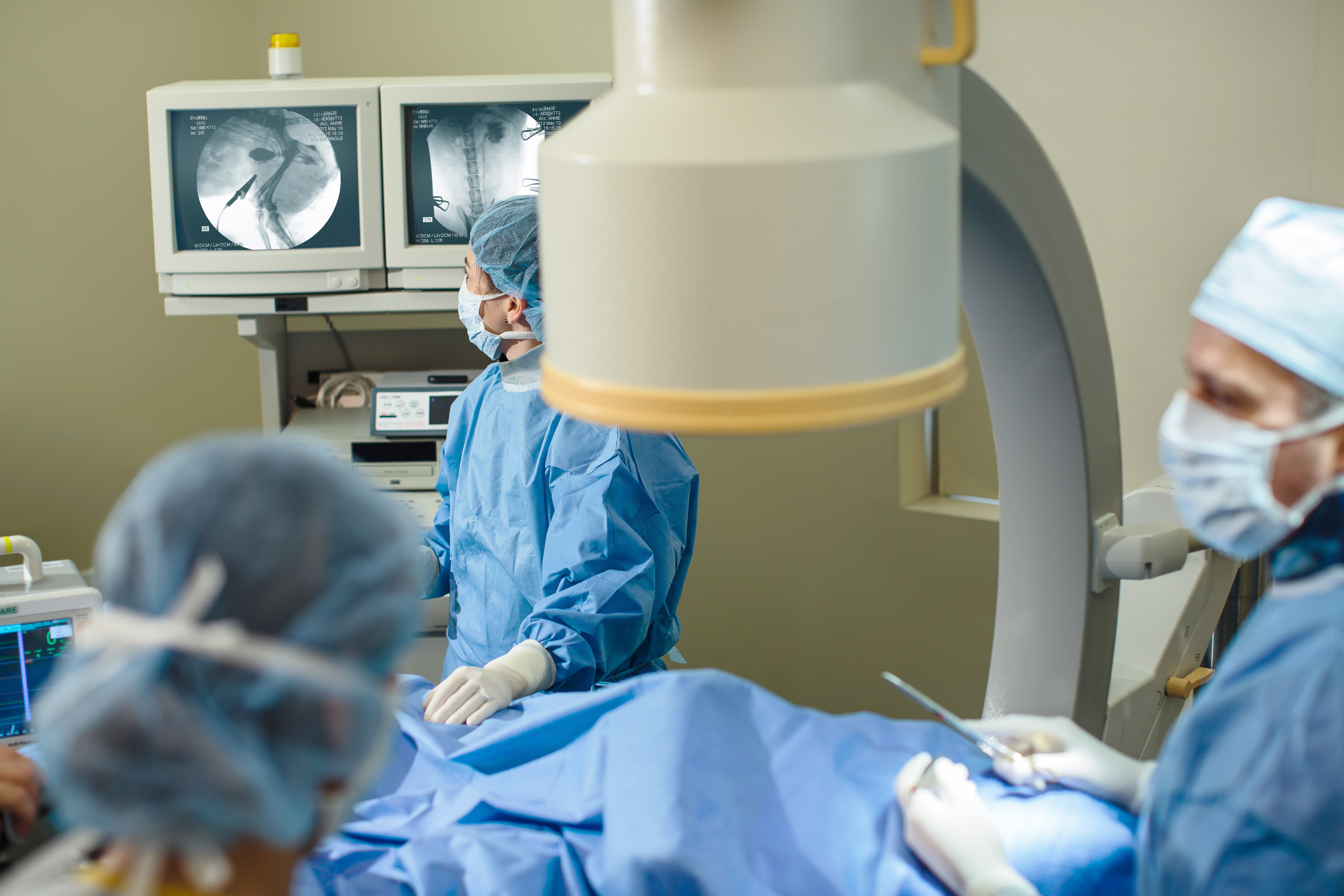
Interventional endoscopy (IE) is the use of endoscopic equipment with other imaging modalities, such as fluoroscopy and/or ultrasound, to perform diagnostic and therapeutic procedures in any part of the body that can be accessed endoscopically (gastrointestinal, biliary, respiratory or urinary tract). This enables access to small orifices and organs that would otherwise require more invasive surgical techniques.
Interventional Radiology and Interventional Endoscopy are well established modalities in human medicine, and have become the standard of care for many conditions in people. The field is far newer in veterinary medicine. CUVS is one of the first and few veterinary centers to offer comprehensive and advanced interventional procedures. In doing so, we can provide treatment options to patients in whom conventional therapies are dangerous or associated with significant morbidity. Moreover, in some cases, these techniques offer treatment options for conditions that simply could not otherwise be treated.
Interventional radiology (IR) is the use of sophisticated imaging modalities, such as fluoroscopy or CT, to gain access to different body structures for diagnostic and/or therapeutic purposes. Similar to its use in human medicine, IR frequently offers non-surgical alternatives, better outcomes and easier recovery, or treatment options for patients with conditions that are not amenable to conventional or other therapy.
Interventional Techniques We Offer
- Tracheal stenting
- Urethral or ureteral stenting
- Nasal stenting
- Stenting of other organs
- Percutaneous cystolithotomy (PCCL)
- Subcutaneous ureteral bypass (SUB)
- Laser lithotripsy or urethral and bladder stones
- Stone basketing and urohydropropulsion
- Laser ablation of ectopic ureters or paramesonephric remnant
- Sclerotherapy for renal hematuria
Conditions We Treat With IR-IE
- Tracheal collapse
- Ureteral or urethral obstruction (via stent placement)
- Kidney, ureteral, and bladder stones
- Obstructive tumors
- Canine incontinence
- Nasal stenosis (narrowing)
- Renal hematuria
Our Specialists
Interventional Radiology and Interventional Endoscopy is performed by CUVS specialists:
Tracheal Stent Placement in a Dog with Collapsing Trachea
Tracheal stent placement is used for the treatment of severe tracheal collapse in patients that have failed medical management. Placement is rapid, usually 30-45 minutes, including initial bronchoscopy, bronchoalveolar lavage, stent sizing and stent placement. View the video of a dog before and after stent placement.
Laser Lithotripsy
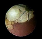 View through an endoscope showing an endoscopically placed retrieval device around a remnant of a bladder stone that had been broken down by laser lithotripsy, enabling endoscopic removal (and avoiding surgery).
 View through an endoscope showing calcium oxalate cystic calculus prior to laser lithotripsy, which is a non-invasive approach for calculus removal.
Urinary Incontinence: Innovative Treatment Options
Urinary incontinence is a relatively common and frequently challenging condition. There are numerous causes, many of which can be managed successfully and non-invasively.
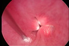
 Urethral sphincter mechanism incompetence (USMI) is common. Cystoscopic-guided urethral bulking agent injections enable us to non-invasively treat these cases. Cystoscopy is used to gain access to the urethra and to identify the region of the urethral sphincter. A bulking agent is then injected into the submucosa of the urethra.
 This is a cystoscopic image of bilateral ectopic ureters.
 In some cases, a hydraulic occluder apparatus is placed around the urethra. The occluder consist of a silicone ring attached to silicone tubing, which connects to a subcutaneous port. Saline is injected into the port to fill the ring, compressing the urethra externally.
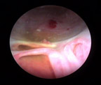
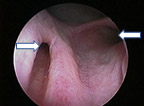 Ectopic ureters are a common cause of urinary incontinence in young dogs. This cystoscopic image is of the urethra of a dog with bilateral ectopic ureters. Arrows show ureters entering the urethra. Cystoscopic laser ablation (CLA) of ectopic ureters is a minimally invasive procedure resulting in a higher success rate than surgery (and a lower complication rate). CLA can be performed at the same time as diagnostic cystoscopy, thus minimizing anesthetic time and cost.
Ureteral Obstructions
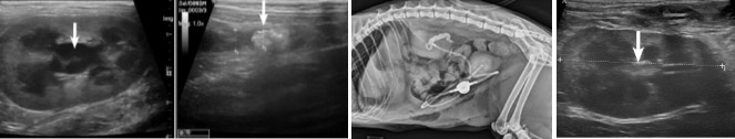
This cat had acute onset vomiting, lethargy, and loss of appetite. The ultrasound reveals left-sided hydronephrosis and hydroureter, with a large hyperechoic structure in the middle. The 3rd image shows placement of a subcutaneous ureteral bypass (SUB) in the left kidney. Note the pigtail of the nephrostomy tube entering the left kidney and the cystostomy tube entering the urinary bladder. The 4th image is 14 days after placement of the SUB system. The left renal pelvis is completely decompressed and the patient’s problems are resolved.